Beckwith-Wiedemannov syndróm
Find a cause you want to help. Every contribution counts.

Všeobecné
Beckwith-Wiedemannov syndróm (skratka BWS, EMG syndrome, anglicky Beckwith-Wiedemann syndrome) nazývaný tiež EMG syndróm (podľa príznakov - Exomphalus, Makroglosia, Gigantizmus) je genetické ochorenie, u ktorého pozorujeme zvýšené riziko vzniku nádorového ochorenia, nadmerného rastu a vrodených malformácií. Príčinou jeho vzniku je zlá génová regulácia (dysregulácia) u génov pre rastový faktor IGF2, ktorý dostávame od otca a génov H19, ktoré naopak získavame od matky. Tento neregulovaný rastový faktor spôsobuje zvýšený rast (gigantizmus) a zvyšuje riziko rastu nádorov. Ochorenie vzniká sporadicky (85%), v 15% sa hlási aj familiárny prenos.
Je pomenovaný podľa Dr. Hansa - Rudolfa Wiedemanna, ktorý prvýkrát opísal súvislosť medzi omfalokélou (vrodená vada postihujúca stenu brušnej dutiny, pri ktorej je prítomná prietrž, do ktorej je vyklenuté črevo), makroglosiou (veľký jazyk) a gigantizmom (veľká dĺžka "koncových" častí tela - nohy, ruky, nos, ...). Neskôr boli popísané ďalšie príznaky prof. Johnom Beckwith, ktorý opísal zväčšenie nadobličiek u osôb s postihnutím, ktoré opísal Wiedemann.
Ochorenie sa vyskytuje s frekvenciou 1: 13 700, kedy je viac viazané na tzv. IVF (umelé oplodnenie). Nepozoruje sa rozdiel vo výskyte u žien či mužov, pričom nie je rozhodujúca ani rasa.
Príznaky:
Pacienti majú tendenciu nadmerného rastu, a to vo zvýšenej miere v priebehu 2. polovice tehotenstva a v prvých rokoch života. Výška doslelých je zvyčajne v normálnom rozmedzí. Abnormálny rast sa môže tiež prejaviť hemihyperpláziou, alebo/a makroglosiou (čo vedie často k ťažkosti pri kŕmení, poruche reči, občas aj spánkového apnoe). Hypoglykémia je hlásená u 30-50% novorodencov.
Medzi ďalšie príznaky patrí omfalokéla, diastáza brušných svalov, embryonálne nádory, prední záhyby na ušnici, visceromegáliu abdominálnych orgánov, fetálnu adrenokortikálnu cytomegáliu (patognomická), renálne (obličkové) abnormality, pozitívna rodinná anamnéza a zriedka rázštep podnebia. Srdcové chyby sa vyskytujú v 9-34% prípadov, asi v polovici prípadov sa pozoruje sponánne odoznievajúca kardiomegália. Kardiomyopatia je zriedkavá. Pacienti sú veľmi náchylní k embryonálnym nádorom, predovšetkým v prvých 8. rokoch života s rizikom okolo 7,5%.
Diagnostika:
Všeobecne platí, že diagnóza je podporovaná prítomnosťou aspoň troch charakteristických klinických nálezov, ale embryonálny nádor sa môže objaviť aj pri "miernejšej" manifestácii. Pozitívne molekulárne testy môžu potvrdiť diagnózu, ale ich negatívny výsledok nevylučuje BWS.
Prenatálne vyšetrenie choriových klkov alebo amniocentézy môžu byť ponúknuté najmä v prípadoch, kedy boli zistené cytogenetické alebo genómove abnormality. Poruchy metylácie sú v súčasnej dobe spoľahlivo detekovateľné pomocou amniocentézy. Amniocentéza môže byť tiež indikovaná pri zachytení nálezov súvisiacich s BWS u plodu počas USG vyšetrenia (napr. fetálna omfalokéla).
Pri chýbaní známeho molekulárneho defektu, môžeme vykonávať skríning s meraním koncentrácie sérového alfa-fetoproteínu u matky a cielené ultrazvukové vyšetrenie u plodu.
Genetické poradenstvo je odporučené, odhad rizika rekurencie a kaskádové genetické testovanie (cascade genetic testing), by mali vziať do úvahy rodinnú anamnézu a molekulárnu podskupinu postihnutého rodinného príslušníka.
Liečba:
Starostlivosť o pacienta zvyčajne zahŕňa štandardnú podpornú lekársku a chirurgickú starostlivosť. Zvýšený dohľad pre riziko vzniku nádorov by sa mal začať, ak je podozrenie alebo je diagnostikovaný BWS a u klinicky nepostihnutého jednovaječného dvojčaťa pacienta, ale momentálne by sa nemal viesť podľa genotypovej /
fenotypovej korelácie. Skríning pre hypoglykémiu by sa mal vykonať v novorodeneckom období, ak existujú predpoklady alebo diagnostické prenatálne nálezy, a to aj u klinicky nepostihnutých novorodencov so zvýšeným rizikom na základe rodinnej anamnézy.
Spektrum klinického priebehu je variabilné, na ťažkom konci spektra sú pacienti ohrození predčasnou smrťou v dôsledku komplikácií vyplývajúcich z hypoglykémie, predčasných pôrodov, kardiomyopatie, makroglosie, alebo vzniku nádorov. U pacientov, ktorí prežijú detstvo je prognóza vo všeobecnosti dobrá.
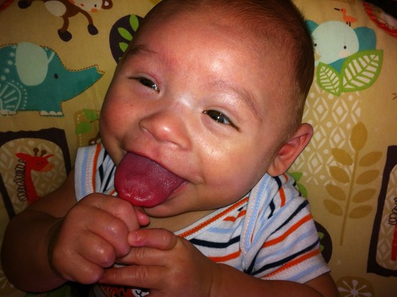
- makroglosia (veľký jazyk)
- makrosómia (veľká pôrodná hmotnosť a dĺžka, ale v dospelosti normálny vzrast)
- defekty brušnej steny (omfalokéla, pupočná prietrž)
- hemihypertrofia (polovica tela je väčšia ako druhá)
- defekty ušných lalôčikov
- neonatálna hypoglykémia (nízka hladina cukru v krvi po pôrode)
- visceromegália - zväčšenie vnútrorných orgánov
- hepatomegália (zväčšenie pečene)
- nefromegália (zväčšenie obličiek)
- ďalej môže byť zväčšená slezina, pankreas, nadobličky
- polyhydramnion (zvýšené množstvo plodovej vody)
- veľká placenta
- anomália obličiek
- Wilmsov tumor (u 5-7% detí s BWS) - tumor obličky vyskytujúca sa u detí
- hepatoblastóm - zhubný nádor postihujúce pečeň u detí
- neuroblastóm - malígny nádor raného detského veku, vychádzajúci z buniek nervového tkaniva
- rhabdomyosarkóm - malígny nádor vychádzajúci z priečne pruhovaného svalu
Pre spojenie sa s ďalšími ľuďmi s rovnakou diagnózou vo vašom okolí sa prihláste.
Prihlásenie