Deficit alfa-1 antitrypsínu
Find a cause you want to help. Every contribution counts.

Všeobecné
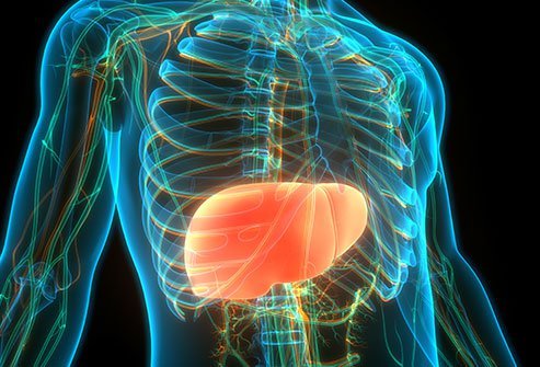
Deficit alfa-1 antitrypsínu (A1AD) je dedičná porucha charakterizovaná nízkymi hladinami proteínu nazývaného alfa-1 antitrypsín (A1AT), ktorý sa nachádza v krvi. Tento nedostatok môže jednotlivca predisponovať k viacerým chorobám a najčastejšie sa prejavuje ako chronická obštrukčná choroba pľúc (vrátane bronchiektázie) a ochorenie pečene (najmä cirhóza a hepatóm) alebo zriedkavejšie ako kožné ochorenie nazývané panikulitída. A1AD je tiež častejší u jedincov s Wegenerovou granulomatózou, teraz nazývanou polyangiitída s granulomatózou.
Nedostatok A1AT umožňuje látkam, ktoré štiepia proteíny (takzvané proteolytické enzýmy), napadnúť rôzne telesné tkanivá. Útok má za následok deštruktívne zmeny v pľúcach (emfyzém) a môže postihnúť aj pečeň a pokožku. Antitrypsín alfa-1 sa bežne uvoľňuje špecializovanými granulami v type bielych krviniek (nazývaných neutrofily alebo polymorfonukleárne leukocyty) v reakcii na infekciu alebo zápal. Nedostatok alfa-1 antitrypsínu má za následok nevyvážený (t.j. relatívne bez odporu) rýchly rozklad bielkovín (aktivita proteázy), najmä v podporných elastických štruktúrach pľúc. Táto deštrukcia môže v priebehu rokov viesť k progresívnemu emfyzému a je urýchlená fajčením, niektorými pracovnými expozíciami a pravdepodobne inými genetickými modifikátormi tohto rizika, ktoré zostávajú neúplne pochopené.
Diagnostika
A1AT je spôsobený mutáciami v géne SERPINA1, ktorý je zodpovedný za produkciu alfa-1 antitrypsínového proteínu. Tento proteín sa normálne produkuje v pečeni a uvoľňuje sa v krvi a funguje tak, že chráni telo pred enzýmom neutrofilnej elastázy. Zdá sa, že A1AT má tiež protizápalové účinky nezávislé od aktivity anti-neutrofilnej elastázy. Mutácie v géne SERPINA1 vedú k produkcii abnormálneho proteínu, ktorý sa zachytí v pečeni, čo má za následok nízke sérové hladiny A1AT, ktoré môžu predisponovať k rozpadu pľúc neutrofilnou elastázou a inými proteolytickými enzýmami (enzýmy, ktoré štiepia proteíny). Abnormálny proteín A1AT sa navyše môže hromadiť v pečeni a spôsobiť poškodenie zjazvením.
Doteraz bolo v géne SERPINA1 identifikovaných viac ako 150 rôznych mutácií, pričom najbežnejší je S a Z, zatiaľ čo normálna verzia (alela) génu sa nazýva M. Alela S spôsobuje, že sérové hladiny A1AT sú stredne nízke a alela Z je spojená s veľmi nízkymi hladinami A1AT v sére (~ 10-15% normálnych hodnôt). Ďalšie vzácne varianty, nazývané nulové, sú spojené s úplnou absenciou A1AT v krvnom obehu, pretože sa nevytvára žiadny proteín.
A1AT je dedičný ako autozomálne ko-dominantné genetické ochorenie. Ko-dominantné genetické poruchy sa vyskytujú vtedy, keď každá dedičná alela vyjadruje určitý účinok (ako znížená hladina A1AT v sére). Všeobecne platí, že v prípade dominantného stavu, keď jedinec zdedí dve kópie abnormálneho génu pre rovnaký znak, jednu od každého rodiča, je riziko ochorenia vyššie, ako keď sa dedí iba jedna abnormálna alela. Ľudia, ktorí majú dve kópie alely Z (ZZ), majú závažný nedostatok A1AT a majú vysoké riziko vzniku emfyzému. Riziko, že dvaja prenášajúci rodičia prejdú zmeneným génom a budú mať postihnuté dieťa (ZZ), je pri každom tehotenstve 25% a za týchto okolností je riziko, že dieťa, ktoré je nositeľom ako rodičia, je 50%. každé tehotenstvo. Nakoniec, šanca, že dieťa dostane normálne gény od oboch rodičov, je 25%. V autozomálnych podmienkach je riziko dedičnosti rovnaké pre mužov a ženy, pretože abnormálny gén sa nenachádza na pohlavných chromozómoch (X alebo Y). V A1AT sa gén SERPIN1A nachádza na dlhom ramene 14. chromozómu.
Ak jedinec dostane jednu normálnu alelu a jednu alelu Z (MZ), klinické riziko vzniku pľúcneho ochorenia sa považuje za malé, aj keď môže existovať podskupina týchto takzvaných heterozygotných pacientov, ktorí sú vystavení vyššiemu riziku, najmä ak sú fajčiari. Ak jedinec dostane jednu alelu S a jednu alelu Z (SZ), považuje sa tiež za zvýšené riziko vzniku chronickej obštrukčnej choroby pľúc, ak fajčí.
Deficit alfa-1 antitrypsínu (A1AD) je porucha, ktorá sa najčastejšie vyskytuje u Severoameričanov alebo u ľudí stredoeurópskeho pôvodu. V Spojených štátoch je diagnostikovaných zhruba 100 000 ľudí s touto diagnózou. Pretože však väčšina prípadov A1AD nie je rozpoznaná, porucha je veľmi málo diagnostikovaná. Odhady naznačujú, že z týchto odhadovaných 100 000 jedincov s vážnym nedostatkom A1AT bolo diagnostikovaných iba u 10% alebo menej, pričom ostatní mali buď chronickú obštrukčnú chorobu pľúc (CHOCHP), ktorá nebola uznaná na základe A1AD, alebo nebola ovplyvnená. Niekoľko dôkazov ukazuje, že A1AD je nedostatočne diagnostikovaný:
1. Mnoho jedincov A1AD zažíva veľké oneskorenia (tj. V priemere 5-8 rokov) medzi počiatočnými príznakmi (často dýchavičnosť) a počiatočnou diagnózou A1AD-
2. Postihnutí jedinci často navštevujú mnohých lekárov so symptómami súvisiacimi s A1AD pred stanovením počiatočnej diagnózy.
3. To, že je naďalej poddiagnostikovaný, naznačuje skutočnosť, že intervaly oneskorenia diagnostiky zostávajú dlhé aj u novšie diagnostikovaných jedincov.
Diagnóza A1AD je založená na nízkej koncentrácii krvnej plazmy A1AT v kombinácii s vysoko rizikovým fenotypom (demonštrovaným izoelektrickým zaostrovaním) alebo genotypom (špecifickou analýzou alel [zvyčajne pre alely Z a S a niekedy aj pre ďalšie alely, ako je napr. Alely F a I a niektoré ďalšie na komerčných testoch]). V niektorých prípadoch je na stanovenie pevnej diagnózy potrebné ďalšie testovanie na sekvenovanie génu A1AT (t.j. mapovanie všetkých chemických prvkov [nazývaných nukleotidy], ktoré tvoria gén A1AT).
Pretože A1AD často nie je rozpoznaný, oficiálne dokumenty s pokynmi odporúčajú, aby boli všetci jedinci s pevnou prekážkou prúdenia vzduchu pri testovaní spirometrie testovaní na poruchu. Tiež by mali byť testovaní všetci priami príbuzní jednotlivcov, u ktorých bola zistená závažná A1AD (t. J. Súrodenci, deti a rodičia), jedinci s panikulitídou a jedinci s nevysvetliteľným ochorením pečene alebo bronchiektáziou.
Je potrebné zvážiť podozrenie na túto poruchu, ak sa emfyzém vyskytne u mladého človeka, nefajčiara alebo niekoho s rodinnou anamnézou emfyzému. Podozrenie na A1AD by malo byť tiež u osôb so žltačkou, hepatitídou, portálnou hypertenziou, hepatocelulárnym karcinómom alebo u osôb s rodinnou anamnézou ochorenia pečene. Ako je uvedené vyššie, poddiagnostikovanie môže vyplývať z testovania iba menšiny rizikových osôb; preto, ako je uvedené vyššie, odporúčania pre testovanie naznačujú, že všetci dospelí so symptomatickou CHOCHP spolu s ďalšími skupinami uvedenými vyššie by mali byť testovaní na A1AD.
Akonáhle je klinické podozrenie na panikulitídu vyvolané sugestívnou anamnézou a fyzikálnym vyšetrením, diagnostikuje sa panikulitída pomocou bioptických vzoriek kožných lézií a krvných testov na stanovenie hladiny cirkulujúceho A1AT a genotypu.
Liečba
Liečba emfyzému spojeného s A1AD zahŕňa štandardné lieky používané na liečbu pacientov s emfyzémom všetkých príčin (ako sú inhalačné bronchodilatátory, inhalačné steroidy, anticholinergiká, kyslíková terapia a podávanie antibiotík alebo inhibítorov fosfodiesterázy 5 na časté respiračné infekcie), ako aj (v špecifických podskupinách) špecifická liečba A1AT nazývaná augmentačná terapia. Cvičebné programy (pľúcna rehabilitácia) a dobrá výživa môžu pomôcť zvýšiť celkovú kvalitu každodenného života. Je veľmi dôležité, aby sa ľudia s emfyzémom vyhýbali fajčeniu, zamestnaniu, ktoré vystavuje pacienta dráždivým látkam pre pľúca, a používaniu nelekárskych aerosólových sprejov. Odporúča sa tiež čo najskôr predchádzať infekcii každoročnou vakcináciou proti chrípke a pravidelným očkovaním proti pneumokokom.
Špecifická liečba A1AD (pre jednotlivcov so zavedeným emfyzémom) môže tiež zahŕňať použitie augmentačnej terapie, ktorá je pravidelnou (zvyčajne raz týždenne) dlhodobou infúziou purifikovaných, združených ľudských ATAT z ľudskej plazmy do žíl s deficitom. V súčasnej dobe šesť liekov na augmentačnú terapiu získalo schválenie od US Food and Drug Administration: Prolastin, Aralast, Aralast NP, Zemaira, Prolastin-C a Glassia, z ktorých posledné štyri sú v súčasnej dobe k dispozícii. Najlepšie dostupné dôkazy naznačujú, že augmentačná terapia môže pomôcť spomaliť progresiu poškodenia pľúc v dôsledku A1AD. Augmentačná terapia nelieči ochorenie pečene súvisiace s A1AD.
U vysoko vybraných pacientov môže byť vhodná chirurgická redukcia objemu pľúc (LVRS) alebo chirurgické odstránenie veľkých splývajúcich oblastí emfyzému (bullae), hoci LVRS môže predstavovať menší prínos pre jednotlivcov s emfyzémom spôsobeným A1AD ako pre jedincov s emfyzémom, ktoré nie sú rozpoznané genetické príčiny. LVRS ako taký sa zriedka odporúča pacientom s A1AD.
Transplantácia pľúc, jednoduchá a dvojitá, bola úspešne vykonaná u mnohých pacientov s A1AD. Táto možnosť liečby sa vykonáva iba u pacientov s ťažkým pľúcnym ochorením v konečnom štádiu, ktorí sa inak kvalifikujú ako kandidáti na takýto chirurgický zákrok.
Na ochorenie pečene súvisiace s A1AD nie je k dispozícii žiadna špecifická terapia, aj keď štúdie na zvieratách preukázali prísľub niekoľkých liekov, ktoré môžu zvýšiť schopnosť pečene odbúravať nevylučovaný A1AT (napr. Rapamycín a karbamazepín), a podnietili výskumné štúdie u jedincov A1AD. Podobne ďalšie prístupy, ktoré sú v súčasnosti predmetom skúmania, zvažujú činidlá, ktoré znížia produkciu abnormálneho proteínu Z pečeňovými bunkami, čo by mohlo predstavovať zníženie rizika pečene, aj keď je však samozrejme potrebné oveľa viac štúdií, než bude možné ponúknuť akýkoľvek záver týkajúci sa týchto súčasných výskumov- založené prístupy. V súčasnej dobe je manažment ochorenia pečene spojeného s A1AD zameraný na kontrolu symptómov. U niektorých ľudí s ochorením pečene spojeným s A1AD môžu byť potrebné špeciálne postupy. Napríklad môžu byť zavedené skratky na zníženie tlaku v cievach v pečeni a rozšírené žily v potravinovej trubici (pažeráku) môžu byť orezané alebo prúžkované, aby sa znížilo riziko krvácania. Transplantácia pečene sa môže odporučiť osobám s terminálnym štádiom ochorenia pečene. Transplantácia normálnej pečene do jedinca s A1AD by mala napraviť abnormality pečene a obnoviť hladiny A1AT v krvi na normálnu úroveň. Transplantácia zároveň prináša určité riziko súvisiace so samotným zákrokom a so zníženou imunitou liekov užívaných na zabránenie odmietnutia transplantovaného orgánu.
Genetické poradenstvo sa odporúča pacientom a ich rodinám.
- pľúcne ochrorenie súvisiace s nedostatkom A1AD (dýchavičnosť, chronický kašeľ, tvorba hlienu, sipot, časté respiračné infekcie, problémy najmä u fajčiarov)
- ochorenie pečene spôsobené A1AD (žltý vzhľad pokožky, zväčšenie pečene, problémy s kŕmením, znížená chuť do jedla, opuch nôh alebo brucha, u dospelých cirhóza, portálna hypertenzia, chronická aktívna hepatitída, hepatocelulárny karcinóm, ospalosť)
- paniulitída - vzácna forma kožného ochorenia
Pre spojenie sa s ďalšími ľuďmi s rovnakou diagnózou vo vašom okolí sa prihláste.
Prihlásenie